You Are Using A 10x Ocular And A 15x Objective
Arias News
Mar 27, 2025 · 5 min read

Table of Contents
Decoding the Microscopic World: Exploring the Power of a 10x Ocular and 15x Objective
The world of microscopy opens up a universe unseen by the naked eye, revealing intricate details of cells, microorganisms, and the minutiae of various materials. Understanding the magnification power of your microscope is crucial to effectively utilizing this powerful tool. This article delves into the implications of using a 10x ocular lens (eyepiece) in conjunction with a 15x objective lens, exploring the resulting magnification, applications, considerations, and limitations of this specific combination.
Calculating Total Magnification: The Simple Equation
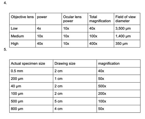
The total magnification of a compound light microscope is determined by multiplying the magnification of the ocular lens by the magnification of the objective lens. In this case, with a 10x ocular and a 15x objective, the calculation is straightforward:
10x (ocular) x 15x (objective) = 150x (total magnification)
This means that the image you observe through the microscope will be 150 times larger than its actual size. This level of magnification is suitable for a wide range of applications, but it's important to understand its limitations compared to higher magnification setups.
Applications of 150x Magnification: What Can You See?
A 150x magnification provides a detailed view of numerous specimens. Here are some common applications:
-
Basic Cell Observation: Viewing the overall structure of plant and animal cells, including the cell wall (in plants), cell membrane, and potentially larger organelles like nuclei and chloroplasts. You won't see the intricate details of smaller organelles at this magnification.
-
Microbial Examination: Observing larger microorganisms like paramecium, amoeba, and some algae. Bacteria will be visible, but discerning their detailed morphology will require higher magnification.
-
Tissue Samples: Examining basic tissue structures. The precise identification of cell types and intricate tissue architecture will benefit from higher magnifications.
-
Prepared Slides: Analyzing pre-prepared slides of various biological and non-biological specimens. This magnification is ideal for educational purposes and for quickly assessing the overall structure of a sample.
-
Simple Material Analysis: Observing the basic structure of fabrics, crystals, or other materials. More detailed analysis of material composition will require specialized microscopes and techniques.
Understanding Resolution: The Limit of Detail
While magnification increases the apparent size of an image, resolution determines the level of detail visible. Resolution refers to the ability to distinguish between two closely spaced points. Even with high magnification, if the resolution is low, the image will appear blurry and lack fine detail.
A 150x magnification setup likely won't have the resolution to clearly distinguish individual organelles within a cell, or to see the fine structures of bacteria. The resolution of a microscope is primarily determined by the numerical aperture (NA) of the objective lens and the wavelength of light. Higher NA values generally indicate better resolution.
Working Distance and Depth of Field: Practical Considerations
The working distance is the distance between the objective lens and the specimen. With a 150x magnification, the working distance will be relatively short, requiring careful focusing and precise positioning of the specimen. This can be challenging, especially for beginners.
The depth of field refers to the thickness of the specimen that is in sharp focus at any given time. At 150x magnification, the depth of field is shallow, meaning that only a very thin layer of the specimen will be sharply focused. This requires meticulous focusing to observe different layers within the sample.
Advantages of Using a 10x Ocular and 15x Objective Combination
Despite the limitations, this combination offers several advantages:
-
Accessibility: Microscopes with this magnification range are relatively common and affordable, making them accessible to students, hobbyists, and researchers with limited budgets.
-
Versatility: Suitable for a broad range of applications, providing a good overview of many different specimens.
-
Ease of Use: Relatively easy to learn and operate, making it a good starting point for beginners in microscopy.
-
Less demanding illumination: Requires less intense illumination compared to higher magnification objectives, minimizing the risk of damaging heat-sensitive specimens.
Limitations and Potential Improvements
While useful, this magnification level has limitations:
-
Limited resolution: Inability to resolve fine details, especially in smaller microorganisms or cellular organelles. For higher resolution, a higher magnification objective lens (e.g., 40x, 100x) with a higher NA would be necessary.
-
Shallow depth of field: Requires precise focusing and can make it difficult to observe three-dimensional structures.
-
Potential for chromatic aberration: At higher magnifications, chromatic aberration (color fringing) can become more noticeable, affecting the clarity and accuracy of the image. Higher quality objectives often mitigate this issue.
Enhancing the Microscopic Experience: Optimization Techniques
To maximize the effectiveness of a 10x ocular and 15x objective combination:
-
Proper Illumination: Use appropriate light intensity and Köhler illumination (if applicable) to ensure even and optimal illumination of the specimen. Too much or too little light can negatively impact image quality.
-
Careful Focusing: Take your time to accurately focus the image, adjusting the fine focus knob carefully.
-
Specimen Preparation: Proper specimen preparation is crucial. For biological samples, staining techniques can enhance contrast and visibility of structures.
-
Immersion Oil (for 100x objectives only): While not applicable to a 15x objective, this section is included for broader microscopic understanding. For higher magnification objectives (100x), immersion oil is typically used to improve resolution and reduce light refraction. This is not necessary and should not be used with lower magnification objectives.
-
Cleanliness: Ensure the lenses are clean and free from dust or debris. Clean lenses are essential for optimal image quality.
Conclusion: A Valuable Tool in the Microscopic Arsenal
The 10x ocular and 15x objective combination offers a practical and accessible entry point into the fascinating world of microscopy. While it may not provide the ultimate resolution for observing subcellular structures or the smallest microorganisms, it serves as a valuable tool for a wide range of applications, particularly for educational purposes, basic cell observation, and the examination of larger microorganisms. Understanding its strengths and limitations allows for its effective use and informs the need for higher magnification setups when greater resolution is required. Remember to always prioritize proper specimen preparation and optimal illumination techniques to get the best possible results from your microscope.
Latest Posts
Latest Posts
-
How Much Is 60 Grams Of Butter
Mar 30, 2025
-
What Is One Overarching Topic Found In Frankenstein
Mar 30, 2025
-
Construction S Symbol With A Vertical Line Through It
Mar 30, 2025
-
What Year Were You Born If You Re 17
Mar 30, 2025
-
How Long Does It Take To Dig A Grave
Mar 30, 2025
Related Post
Thank you for visiting our website which covers about You Are Using A 10x Ocular And A 15x Objective . We hope the information provided has been useful to you. Feel free to contact us if you have any questions or need further assistance. See you next time and don't miss to bookmark.
